What Is the Functional Unit of a Muscle Cell
Specialized Cell Muscle Cell. The chemical in every cell that carries genetic information.

Actin Wikipedia Muscle Contraction Medical School Stuff Biology
Structure edit edit source Myocytes can be incredibly large Diameters of up to 100 micrometers.
. The functional contractile unit of the myofibril of a striated muscle. Functional resistance training methods for targeting patient-specific gait deficits. The inner endocardium lines the cardiac chambers covers the cardiac valves and joins with the endothelium that lines the.
The sarcomere is the basic contractile unit of skeletal muscle. March 18 2022. It has a centrally placed nucleus.
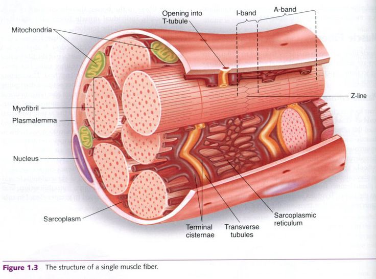
Sarcomeres are contractile units of skeletal muscle that divide into I and A bands M and Z lines and the H zone. The length of the sarcomere varies with muscle activation. This rest length is less than the thick filament.
A review of devices and their effects on muscle activation neural control and gait mechanics. This muscle assists in breathing. These fibers are the functional unit of the organ leading to contraction and relaxation.
To understand how a muscle contracts you need to know a bit about. In contrast muscle fibers making up the stapedius a small muscle of the inner ear are only a few millimeters in length. The myofibrils consist of thousands of these sarcomeres strung end to end.
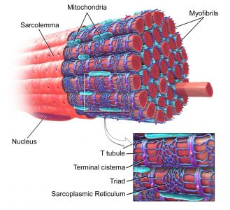
A single muscle fiber is composed mostly of actin and myosin fibers covered by a cell membrane sarcolemma. Muscle fibers which form contractile unit of heart 99 ii. There are two major classifications of skeletal muscle.
Thick filaments are organized bundles of myosin while thin filaments are made of actin along with the two. Type I slow oxidative and Type II fast-twitch. So impulses travel along muscle cell membranes just as they do along nerve cell membranes.
This is essentially a tube that carries various bodily fluids. Muscle fibers which form conductive system 8 9. The pancreatic duct runs the full length of the pancreas and drains into the duodenum.
Sarcomere of the cardiac muscle has all. Cardiac muscle tissue or myocardium forms the bulk of the heart. Muscle fibers which form pacemaker iii.
In the anterior thigh a muscle fiber may be a meter long. However the function of impulses in muscle cells is to bring about contraction. A small anatomic structure.
The muscle cell that makes up animal skeletal muscle is a classic example of a syncytium cell. The functional unit of contraction or force production is the sarcomere extending from one Z-disk to the next. Typically the rest length is about 2022 μm depending on the length of the thin filament.
The term may also refer to cells interconnected by specialized membranes with gap junctions as seen in the heart muscle cells and certain smooth muscle cells which are synchronized electrically in an action potential. A dome shaped muscle that separates the lungs and heart from the abdomen. Skeletal muscle fibers can be quite large for human cells with diameters up to 100 μm and lengths up to 30 cm 76 in in the Sartorius of the upper legDuring early development embryonic myoblasts each with its own nucleus fuse with up to hundreds of other myoblasts to form the.
Because skeletal muscle cells are long and cylindrical they are commonly referred to as muscle fibers. Clinical Biomechanics is an international multidisciplinary journal of biomechanics with a focus on. In cultured skeletal muscle cells and C2C12 myotubes Ucn 2 inhibits insulin-induced Akt and ERK12 phosphorylation consistent with the hypothesis that Ucn 2 functions as a local negative regulator of glucose uptake in skeletal muscle and suggests the possibility that suppression of the Ucn 2CRH-R2 pathway may provide benefits in insulin.
Myofibrils are embedded in the sarcoplasm. The impetus of the membership remains research-based academic surgery and to promote the shared vision of research and academic pursuits through the exchange of ideas between senior surgical residents junior faculty and established. The Sarcoplasm Rich with.
The cell membrane of a muscle cell is called the sarcolemma and this membrane like that of neurons maintains a membrane potential. The Association for Academic Surgery is widely recognized as an inclusive surgical organization. The heart wall is a three-layered structure with a thick layer of myocardium sandwiched between the inner endocardium and the outer epicardium also known as the visceral pericardium.
Striated and resemble the skeletal muscle fibre Cardiac muscle fibre is bound by sarcolemma. The vast diversity in the makeup of skeletal muscle leads to. It is made of thick and thin filaments.
About the Societies.
Muscle Cell Myocyte Definition Function Structure Biology

Associative And Tangental Energetic And Emotional Signal Bundle That Is Mirrored In The Physical World Human Anatomy And Physiology Physiology Muscular System

Cumulative Topic 6 Microanatomy Of Myofiber Body Muscle Anatomy Skeletal Muscle Anatomy Muscle Anatomy
Myofibrils Complete Soccer Training Functional Anatomy Of The Skeletal Muscle Muscle Muscle Anatomy Skeletal Muscle Anatomy


Comments
Post a Comment